The Central Nervous System
Dr. C. George Boeree
The Central Nervous System
Dr. C. George Boeree
This page has been translated into Swedish by Eric Karlsson
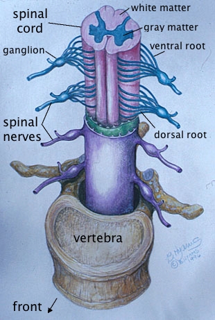
The spinal cord runs from the base of the skull all the way down the
spine to
the "tail bone." The neurons are found in an H-shaped space
within
the spinal vertebrae. There are motor pathways coming down from
the
brain and sensory pathways going up to the brain. Sensory nerves
enter into the back parts (dorsal roots) of the "H," while motor
neurons exit the forward parts (ventral roots) of the "H."
Interneurons often connect these sensory and motor neurons.
Besides sending messages up and down to and from the brain, the spinal cord has another very important function: Reflexes. In fact, in very simple animals, that is the main function of the cord. Basically, a reflex is the connection of sensory neurons, via interneurons, to motor neurons. For example, there are pain sensors in your fingers. If you hold your finger over a flame for a period of time, the pain will trigger motor neurons to pull your finger away. It is true that you can over-ride this reflex with "will power," but as the example intentionally shows, it isn't easy!
Reflexes do much more than just get your finger out of the fire: A great deal of movement is accomplished through reflexes. Even brand new babies already have the necessary reflexes for walking: If you hold a baby and gently touch its feet to the floor, it will start making step-like movements! All that is needed is the muscle strength to stand and, of course, a lot of practice.
The Brain
The brain is traditionally divided into three parts, the hindbrain,
the midbrain, and the forebrain. This drawing is roughly what it
would look like if you sliced your brain straight down the middle, like
a part in your hair. The front of the brain is on the left, the
back on the right:
The hindbrain or brain stem consists of three parts. The first is the medulla, which is actually an extension of the spinal cord into the skull. Besides containing tracts up and down to and from the higher portions of the brain, the medulla also contains some of the essential nuclei that govern respiration and heart rate. The upper part of the medulla contains a pinky-sized complex of nuclei called the reticular formation. It is the regulatory system for sleep, waking, and alertness.
The second part is the pons, which means bridge in Latin. The pons sits in front of the medulla, and wraps around it to the back. It is primarily the pathways connecting the two halves of the next part, which is called the cerebellum.
The cerebellum, which means "little brain" in Latin, is in fact shaped like a small brain, and it is primarily responsible for coordinating involuntary movement. It is believed that, when you learn complex motor tasks, the details are recorded in the cerebellum.
The midbrain is, in human beings, the smallest part of the brain. It connects the hindbrain to the forebrain, and contains several pathways important to hearing and vision. It is much larger in lower animals and in the human fetus.
The largest and, for psychologists, most interesting part of the brain is the forebrain. It starts with the thalamus, which is practically in the center of your head. The thalamus is like a switching station, conducting signals from the body up to the relevant parts of the higher brain, and down from the brain to the lower brain and spinal cord.
The forebrain, though, is complex enough to require its own chapter -- two, in fact: One for the limbic system, and one for the cerebrum.
