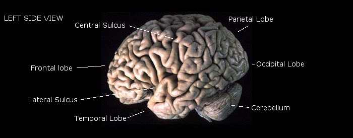
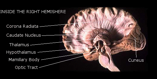
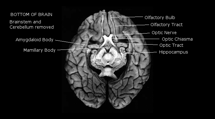
Images from The Virtual Hospital
http://www.vh.org/Providers/Textbooks/BrainAnatomy/BrainAnatomy.html
(all labeling errors my own!)


| Before the 20th century, there
was only one way to see the brain: Open up the skull. Of
course, with the assistance of a talented medical artist or, later, a
good photograph, this provides us with a particularly good view.
For the most part, however,
it required a dead patient! Fortunately, we now have a number of
imaging
techniques that allow us to see what is going on inside the brain of a
living
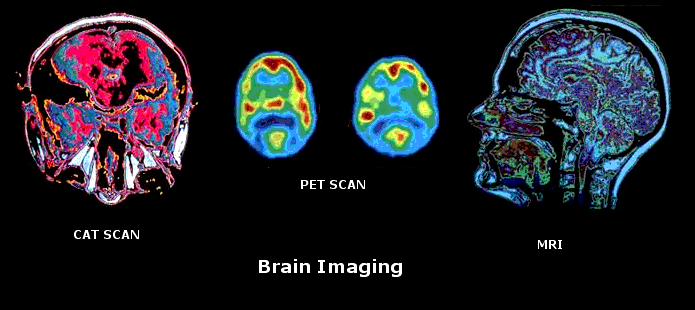
human being. X-rays were the first things used to look at a living brain. While some details are visible, the nature of the brain is such that it is not a particularly good subject for the X-ray. The CT scan (computer tomography) or CAT scan involves taking a large series of x-rays from various angles, and then combining them into a three-dimensional record on a computer. The image can be displayed and manipulated on a computer screen. The PET scan (positron emission tomography) works like this: The doctor injects radioactive glucose ("sugar water") into the patient’s bloodstream. The device then detects the relative activity level - that is, the use of glucose - of different areas of the brain. The computer generates an image that allows the researcher to tell which parts of the brain are most active when we perform various mental operations, whether it’s looking at something, counting outloud, imagining something, or listening to music! The MRI (magnetic resonance imaging) works like this: You create a very strong magnetic field which runs through the person from head to toe. This causes the spinning hydrogen atoms in the person’s body to line up with the magnetic field. Then you send a radio pulse at a special frequency that causes the hydrogen protons to spin in a different direction. When you turn off the radio pulse, the protons will return to their alignment with the magnetic field, and release the extra energy they took in from the radio pulse. That energy is picked up by the same coil that produced the energy, now acting like a three dimensional antenna. Since different tissues have different relative amounts of hydrogen in them, they give a different density of energy signals, which the computer organizes into a detailed three-dimensional image. This image is nearly as detailed as an anatomical photograph! © Copyright 2003, 2009 C. George Boeree |
